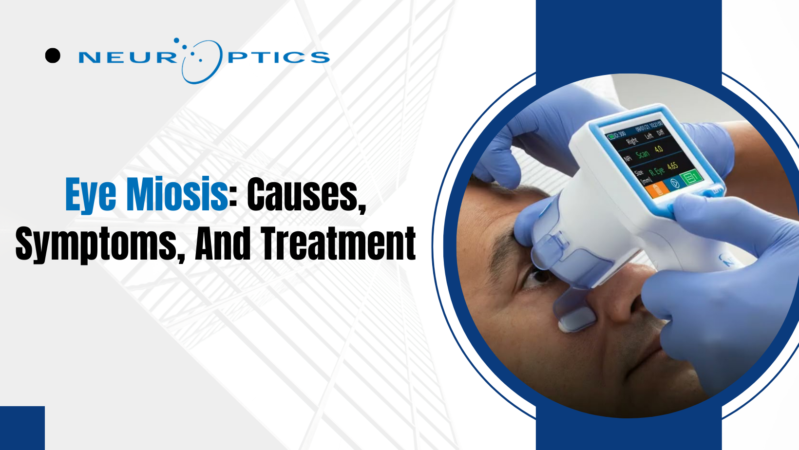
Eye Miosis: Causes, Symptoms, And Treatment
Introduction
The Iris is known as the diaphragm of the eye. The aperture on the Iris through which light enters the human eye is the pupil, which dilates or constricts depending upon light entering the eye.
The diameter of the pupil is usually 2-4 mm in size. The constriction or a reduction of the pupil’s diameter is known as eye miosis.
You should note that the human pupil works like the aperture of a lens used in cameras. The aperture is designed to increase or decrease to allow the passage of light. Similarly, your pupils dilate in the absence of light and constrict in the presence of it.
However, this natural response to external stimulation is often disrupted, and your eyes can exhibit abnormal miosis. This is caused by various reasons, including trauma, tumors, and infections. It is also rendered as a side effect of multiple drugs or exposure to certain chemicals.
Pathway of Light and Pupillary Light Reflex
The retina is stimulated when light falls on your eyes, and the optic nerve carries an impulse to the brain where the pretectal nucleus in the midbrain is stimulated. This requires a further bilateral stimulation of the third cranial nerve nucleus.
An efferent from this third cranial nerve constricts the bilateral constrictor muscles in the Iris of both the eyes, which in turn leads to miosis. This process of pupillary light reflex, also known as the light reflex pathway, is crucial to human vision.
However, the pupillary response in traumatic brain injury and the causes mentioned above can lead to prolonged miosis in one or both your eyes, and such a condition may require a consultation with an ophthalmologist.
Causes of miosis and the evaluation of pupillary reactions associated with this specific medical condition
As explained before, miosis is a natural phenomenon observed in the human eye. It can be caused as a result of
● A response to intense light
● Sleep! Your pupils usually are miotic when you sleep; this is often referred to as a pinpoint.
However, as mentioned above, a more critical situation leading to prolonged miosis may arise for the following reasons.
Read on to get comprehensive insights into these causes and the extent of miosis.
Pontine hemorrhage
The pons in your mid-brain can suffer a hemorrhage that can lead to a pinpoint pupil due to hypertension. You should lookout for a sudden increase in body temperature as this condition is often accompanied by a high fever.
Traumatic brain injury
Neurological damage or injury to the head causes miosis. Prolonged miosis confirmed by the pupillary response in traumatic brain injury is found in patients involved in severe accidents. The pupils constrict and are often unresponsive to external stimuli.
The side effect of drugs
Parasympathomimetic drugs like methacholine, bethanechol, physostigmine, etc., are known to have caused eye miosis. Side effects of these drugs alter the ability of your pupils to dilate in the absence of light.
Exposure to chemicals and their effects on pupil reactivity
Pesticides that contain organophosphate groups and carbamates can cause miosis too. Prolonged exposure to these chemicals leads to the loss of effectiveness of the bilateral constrictor muscles, and pupil reactivity suffers.
Iridocyclitis
This is a condition that is characterized by narrow and non-reactive pupils. Caused mainly by infections, pupillary size measurement can reveal pupils of irregular size.
Pupil evaluation and its role in Horner’s Syndrome
Horner’s Syndrome is mainly reported to have been unilateral, i.e., observed in only one eye is primarily a result of injuries, birth trauma, or even a lung tumor known as Pancoast tumor.
An individual suffering from Horner’s Syndrome will exhibit the following symptoms.
1. Abnormal drooping of the upper eyelids.
2. Loss of ciliospinal reflex.
3. Pseudo enophthalmos that causes the eyes to appear larger than usual.
Interestingly, chronic miosis can be observed in older adults too. This condition is referred to as Senile Rigid Miotic Pupil in the medical community.
Diagnosis of Eye Miosis through the evaluation of pupillary reaction to external stimuli
The critical examinations to diagnose an individual with eye miosis are as follows.
Direct light reflex to determine pupil reactivity
You will be examined in a dimly lit room. Upon being asked to cover an eye with your palm, the physician shall throw light at the other eye to evaluate pupil reactivity.
A similar process is carried out for the other eye as well. Normal pupil reaction will show brisk constriction to light, and the constriction is maintained during the entire period of exposure to light.
Consensual light reflex for the evaluation of pupillary reaction
A dimly lit room is required for this examination as well. A partition (often cardboard) shall be placed between your eyes to separate them. One eye is then exposed to light, and an evaluation of pupillary reaction in the other eye is observed.
Near reflex
When we look at something kept close to our face, the eyes converge, and the pupils constrict. This results from the accommodation reflex associated with bilateral pupil constriction, convergence, and accommodation.
You shall be asked to focus on an object placed roughly 15 cm away, and your pupil reactivity shall be examined.
Swinging flashlight test
This test helps reveal any afferent defect in your eyes. A flashlight is aimed at one pupil, and constriction is noted. Following this, the light is quickly moved to the other eye to observe its constriction.
This incessantly rapid to and fro movement is executed several times to determine the current state of the pupil as well as pupil reactivity to external stimuli.
Treatment of Eye Miosis to improve pupil reactivity
A positive diagnosis of eye miosis shall require swift action. It is important to note that eye miosis is often a symptom that is an indicator of other underlying health conditions that dramatically affect pupil reactivity.
To successfully treat miosis, it is crucial to determine the reason behind miosis and treat those symptoms. If medication is determined to be the reason behind this condition, your physician will suggest a different class of drugs.
Miosis caused by pontine hemorrhage is treated using antihypertensives, which are instrumental in bringing your blood pressure down.
A craniotomy is suggested for miosis caused by a hematoma. Your physician shall ask you to follow up with a cranioplasty after 6-12 months of craniotomy.
However, cycloplegics like atropine or homatropine are also used to treat miosis that is caused as a result of iridocyclitis. These medicines are used to dilate your pupil, and continued use shall alleviate your symptoms.
A prompt examination and subsequent treatment are essential to restore the normal functions of your pupils.
Conclusion
Eye Miosis is a condition that is primarily found in individuals suffering neurological trauma or from certain health conditions like iridocyclitis, Horner’s Syndrome, etc. An abnormal and prolonged constriction of the pupil is observed following diagnostic tests for the evaluation of pupillary reaction.
Your pupillary light reflex suffers and can lead to permanent eye damage. It is advised that you visit your physician immediately since this condition is required to be treated by a licensed ophthalmologist.

Post Comment